Presentation
Aphthous ulcers, also known as canker sores, present as single or multiple ulcerations in the oral mucosa. The first onset of these benign lesions usually occurs in childhood or early adulthood. Canker sores affect women more than men, are self-limiting, and are disproportionately painful relative to the size of the lesion. Because of the pain severity these ulcers can cause, patients typically present to the dental office worried about malignancy and looking for treatment. It is important that the clinician be well-informed about aphthous ulcers and their treatment in order to alleviate both patient concerns and pain.
Types of canker sores
There are three major categories of aphthous ulcers (canker sores):1
- 80% of canker sores are minor aphthous ulcers. These appear as round white lesions 2 mm to 8 mm in size, surrounded by a thin red halo usually occurring on nonkeratinized oral mucosa with a duration time of 10 to 14 days (figure 1).
- 10% to 15% of canker sores are major aphthous ulcers, usually larger than 10 mm in diameter, and can occur on both keratinized and nonkeratinized mucosa. These lesions can last two to eight weeks and may cause scarring in some cases (figure 2).
- 5% to 10% of canker sores are herpetiform aphthous ulcers, which are multiple ulcerations 2 mm to 3 mm in size that, when close enough to each other, can coalesce into one large ulcer. These types of canker sores are found on keratinized and nonkeratinized oral mucosa, last 10 to 14 days, and can be very painful (figure 3).
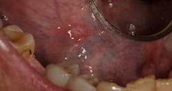
Figure 1: Minor aphthous ulcer
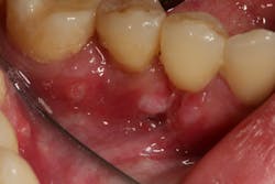
Figure 2: Major aphthous ulcer
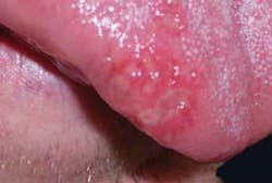
Figure 3: Herpetiform aphthous ulcer (Photo courtesy of Lisa Dowst-Mayo, RDH, BSDH)
Etiology and known triggers
Canker sore attacks can be brought on by a variety of factors including stress, medications, hormonal changes, vitamin deficiency (especially B12, folic acid, and iron), sensitivities to foods, and dental ingredients, such as sodium lauryl sulfate found in toothpaste. In addition, trauma from dental procedures can precipitate an attack, leading the patient to believe that the dental treatment itself was the cause of the aphthous ulcer. Genetics, immunologic factors, and microbial factors also play a role in recurrent aphthous ulcers.2
Diagnosis and associated systemic disorders
Diagnosis is based on clinical presentation along with medical history information. Once an aphthous ulcer is identified, it is important for the dentist to thoroughly review the patient’s medical history for possible systemic disease. Associated disorders include gastrointestinal disorders, such as celiac disease and inflammatory bowel disease; hematologic disorders, such as anemia; nutritional deficiency; and immunologic disorders, such as Behçhet's syndrome or HIV/AIDS. Important note: If the lesion does not resolve in two weeks, further screening such as blood tests may be warranted to rule out these systemic disorders.
Treatment of canker sores often takes a "wait until it goes away" approach or can be directed at providing pain relief. Over-the-counter analgesics containing benzocaine, such as Orajel/Orabase, are often used. Antimicrobial mouth rinses, such as .12% chlorhexidine, can also be prescribed.3
New treatment options
The following treatment has been shown to decrease severity of pain and duration of more painful canker sore lesions:
1. Magic mouthwash rinse: Although there are many different formulations, the typical magic mouthwash prescription includes one-part viscous lidocaine, one-part diphenhydramine, and one-part Maalox/Mylanta or alternative. This combination of ingredients numbs the oral mucosa and decreases inflammation to reduce pain. The Maalox helps preserve the other ingredients and provides a protective coating to the canker sores. Magic mouthwash can also include tetracycline as well to prevent the sores from growing larger (an option is to add a steroid such as prednisone/dexamethasone and an antifungal such as nystatin).4
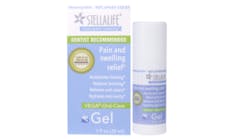
2. StellaLife Products: Homeopathic remedies utilizing essential oils and other anti-inflammatory agents in mouth rinses have shown efficacy in treating aphthous ulcers.5 For aphthous ulcer treatment, StellaLife offers a gel (figure 4) that is applied to the ulcer area with a clean cotton swab five to seven times a day. This application can decrease pain intensity and reduce ulcer duration from 10 to 14 days down to just two to three days after onset. The gel’s active ingredients include Aconitum, Azadirachta, Arnica, Calendula, Chamomilla, Echinacea, Gelsemium, Hepar, Hypericum, Ignatia, Mercurius, Plantago, and Ruta. This ingredient combination contains anti-inflammatory properties that are shown to accelerate healing, temporarily relieve pain, and reduce swelling and bruising.
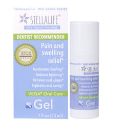
Figure 4: StellaLife VEGA Oral Care Gel relieves pain associated with canker sores and reduces ulcer duration.
3. Laser ablation therapy: Studies have shown that laser treatment can help to reduce the duration and intensity of aphthous ulcers.6 Lasers can be very effective in cases of major aphthous ulcers and herpetiform aphthous ulcers, where pain intensity can be high and the patient is looking to reduce lesion duration and scarring potential. In a recent case series, a 9.3-micron CO2 laser demonstrated efficacy in the treatment of aphthous ulcers. The area of the canker sore is anesthetized with local anesthesia, and the lesion is ablated for 10 to 20 seconds (figure 5). This treatment has been shown to reduce both the duration and intensity of the canker sore lesion by 50% to 75%.7
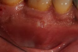
Figure 5: Laser ablation therapy of an aphthous ulcer
References
1. Altenburg A, El-Haj N, Micheli C, Puttkammer M, Abdel-Naser MB, Zouboulis CC. The treatment of chronic recurrent oral aphthous ulcers. Dtsch Ärztebl Int. 2014;111(40):665-673. doi: 10.3238/arztebl.2014.0665.
2. Preeti L, Magesh KT, Rajkumar K, Karthik R. Recurrent aphthous stomatitis. J Oral Maxillofac Pathol. 2011;15(3):252-256. doi:10.4103/0973-029X.86669.
3. Belenguer-Guallar I, Jiménez-Soriano Y, Claramunt-Lozano A. Treatment of recurrent aphthous stomatitis. A literature review. J Clin Exp Dent. 2014;6(2):e168-e174. doi: 10.4317/jced.51401.
4. Magic Mouthwash. Pharmacist’s Letter/Prescriber’s Letter 2007;23(7):230703.
5. Hoseinpour H, Peel SA, Rakhshandeh H, et al. Evaluation of Rosa damascena mouthwash in the treatment of recurrent aphthous stomatitis: a randomized, double-blinded, placebo-controlled clinical trial. Quintessence Int. 2011;42(6):483-491.
6. Tezel A, Kara C, Balkaya V, Orbak R. An evaluation of different treatments for recurrent aphthous stomatitis and patient perceptions: Nd:YAG laser versus medication. Photomed Laser Surg. 2009;27(1):101-106. doi: 10.1089/pho.2008.2274.
7. Froum SH, et al. The treatment of recurrent aphthous ulcers using a 9.3-micron CO2 laser, a case series manual in preparation.

Scott Froum, DDS, a graduate of the State University of New York, Stony Brook School of Dental Medicine, is a periodontist in private practice at 1110 2nd Avenue, Suite 305, New York City, New York. He is the editorial director of Perio-Implant Advisory and serves on the editorial advisory board of Dental Economics. Dr. Froum, a diplomate of the American Board of Periodontology, is a volunteer professor in the postgraduate periodontal program at SUNY Stony Brook School of Dental Medicine. Contact him through his website at drscottfroum.com or (212) 751-8530.
About the Author

Scott Froum, DDS
Editorial Director
Scott Froum, DDS, a graduate of the State University of New York, Stony Brook School of Dental Medicine, is a periodontist in private practice at 1110 2nd Avenue, Suite 305, New York City, New York. He is the editorial director of Perio-Implant Advisory and serves on the editorial advisory board of Dental Economics. Dr. Froum, a diplomate of both the American Academy of Periodontology and the American Academy of Osseointegration, is in the fellowship program at the American Academy of Anti-aging Medicine, and is a volunteer professor in the postgraduate periodontal program at SUNY Stony Brook School of Dental Medicine. He is a trained naturopath and is the scientific director of Meraki Integrative Functional Wellness Center. Contact him through his website at drscottfroum.com or (212) 751-8530.