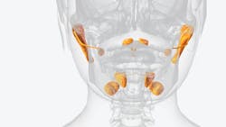
The tubarial salivary glands: A newly recognized organ with dental significance
What you'll learn in this article
- How the tubarial salivary glands were recently discovered using advanced PSMA PET/CT imaging
- Why these glands are important for maintaining nasopharyngeal lubrication, swallowing, and mucosal health
- The role of tubarial glands in radiation therapy planning and their impact on xerostomia and oral health outcomes
- Broader clinical implications for dentistry, oncology, and interdisciplinary care in preserving salivary function
For over a century, anatomy textbooks have described three paired major salivary glands—the parotid, submandibular, and sublingual. Beyond these, hundreds of minor salivary glands scattered throughout the oral cavity have been recognized. Yet in 2020, radiologists and oncologists using advanced PSMA PET/CT imaging discovered a previously overlooked structure: the tubarial salivary glands, located in the nasopharynx near the torus tubarius.1 Their recognition has profound implications for dentistry, oncology, and head and neck medicine.
Why have the tubarial glands just been discovered?
The tubarial glands escaped detection until recently because:
- Location: They lie deep in the nasopharyngeal tissue, an area difficult to visualize with conventional imaging and endoscopy.
- Technology: Standard CT, MRI, and traditional histologic dissection often failed to identify this tissue as distinct from surrounding mucosa. The development of prostate-specific membrane antigen (PSMA) PET imaging, used primarily in prostate cancer detection, provided the high sensitivity needed to highlight glandular tissue.
- Assumptions: Anatomists historically assumed all salivary tissue was accounted for, leaving little incentive to investigate hidden regions.
This combination of technological limitations and confirmation bias explains why the glands were only described in the last five years.
Why are tubarial salivary glands important?
The tubarial glands appear to play a critical role in lubricating and protecting the nasopharyngeal and oropharyngeal region.2 They secrete mucous and serous fluid that contribute to:
- Moistening the pharyngeal walls
- Facilitating swallowing
- Reducing friction during speech and respiration
- Maintaining mucosal health in an area often exposed to irritants
In short, these glands provide a unique contribution to the upper aerodigestive tract’s hydration and function.
Significance in radiation treatment
For patients undergoing radiation therapy for head and neck cancers, preservation of salivary gland tissue is vital. Xerostomia (dry mouth) is one of the most debilitating complications of radiation, affecting speech, swallowing, taste, oral microbiome balance, and quality of life.
Until recently, radiation treatment planning did not account for the tubarial glands. This omission may have inadvertently increased collateral damage to these glands, compounding the risk of:
- Severe xerostomia
- Dysphagia (difficulty swallowing)
- Mucosal infections and increased caries risk
- Reduced patient compliance and poorer oncologic outcomes
Recognizing the tubarial glands allows oncologists to redefine target volumes and adapt radiation therapy planning, sparing this tissue when possible.
Keeping the mouth moist and functional
Unlike the parotid and submandibular glands that deliver saliva directly into the oral cavity, the tubarial glands hydrate the nasopharyngeal corridor—a crucial upstream location for overall oral moisture.3 Protecting them could mean less chronic dryness, better mucosal resilience, and fewer long-term complications post-radiation.
This is particularly significant because xerostomia is often irreversible once salivary glands are destroyed. Awareness of the tubarial glands creates new opportunities for saliva-sparing therapies and proton beam targeting protocols.
The broader significance of their discovery
The discovery of the tubarial glands is a reminder that even in 2025, human anatomy is not fully mapped.4 For health professionals, it reinforces several key points:
- Anatomy evolves with technology: New imaging can redefine what we think we know.
- Multidisciplinary awareness is critical: Dentists, oncologists, ENTs, and radiologists must collaborate when adapting treatment plans.
- Patient quality of life matters: Protecting salivary function is not cosmetic—it directly impacts nutrition, oral health, and overall well-being.
Conclusion
As more research emerges, the tubarial glands may redefine best practices in head and neck oncology, salivary gland preservation, and oral-systemic health management. The tubarial salivary glands represent a newly described structure with profound clinical implications. Their discovery underscores the importance of precision medicine, careful radiation planning, and the ongoing evolution of our anatomical understanding.
Editor’s note: This article originally appeared in Perio-Implant Advisory, a chairside resource for dentists and hygienists that focuses on periodontal- and implant-related issues. Read more articles and subscribe to the newsletter.
References
- 1. Valstar MH, de Bakker BS, Steenbakkers RJHM, et al. The tubarial salivary glands: a potential new organ at risk for radiotherapy. Radiother Oncol. 221;154:292-298. doi:10.1016/j.radonc.2020.09.034
- 2. Ling SW, van der Veldt A, Segbers M, Luiting H, Brabander T, Verburg F. Tubarial salivary glands show a low relative contribution to functional salivary gland tissue mass. Ann Nucl Med. 2024;38(11):913-918. doi:10.1007/s12149-024-01965-x
- 3. Pringle S, Bikker F, Vogel W, et al. The tubarial glands resemble the palatal salivary glands based on further histological characterization, and may represent an organ of interest in primary Sjögren’s syndrome [abstract]. Arthritis Rheumatol. 2022;74(suppl 9).
- Ebrahim A, Reich C, Wilde K, Salim AM, Hyrcza MD, Willetts L. A comprehensive analysis of the tubarial glands. Anat Rec (Hoboken). 2025;308(5):1425-1437. doi:10.1002/ar.25561
About the Author

Scott Froum, DDS
Editorial Director
Scott Froum, DDS, a graduate of the State University of New York, Stony Brook School of Dental Medicine, is a periodontist in private practice at 1110 2nd Avenue, Suite 305, New York City, New York. He is the editorial director of Perio-Implant Advisory and serves on the editorial advisory board of Dental Economics. Dr. Froum, a diplomate of both the American Academy of Periodontology and the American Academy of Osseointegration, is in the fellowship program at the American Academy of Anti-aging Medicine, and is a volunteer professor in the postgraduate periodontal program at SUNY Stony Brook School of Dental Medicine. He is a trained naturopath and is the scientific director of Meraki Integrative Functional Wellness Center. Contact him through his website at drscottfroum.com or (212) 751-8530.
