Postextraction hemostasis: An updated protocol for managing the anticoagulated dental patient
Key Highlights
- Don’t stop anticoagulants by default: For most routine extractions, meticulous surgical technique and local hemostatic measures are safer than interrupting anticoagulation therapy.
- Hemostasis starts in the operatory: Complete granulation tissue removal, atraumatic surgery, socket packing, primary closure, and adequate pressure are the strongest predictors of successful bleeding control.
- Preparation prevents emergencies: Know the patient’s anticoagulant, coordinate with their physician when appropriate, and provide clear postoperative instructions to minimize after-hours bleeding complications.
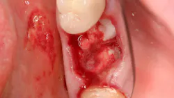
One of the most anxiety-inducing phone calls a clinician can receive comes after hours: a patient you extracted teeth from earlier that day is bleeding and cannot get it to stop. If that patient is on anticoagulation medication, the anxiety compounds quickly. But here is the clinical reality most practitioners miss: the medication is rarely the primary problem. Inadequate surgical technique almost always is.
The reflex to simply discontinue anticoagulation before every extraction is not only outdated but potentially dangerous. Stopping a patient’s Coumadin or Eliquis for a minor procedure introduces a real and measurable risk of thromboembolic events: stroke, pulmonary embolism, and myocardial infarction. What the literature now supports is clean surgery combined with appropriate local hemostatic measures is sufficient for the vast majority of anticoagulated patients undergoing routine extractions.1 The question is not whether to operate on these patients. The question is whether your technique and your protocol are good enough.
Why patients are anticoagulated: Know before you cut
Before any surgical intervention, you must understand why your patient is anticoagulated. The most common indications include atrial fibrillation, mechanical heart valves, deep vein thrombosis, pulmonary embolism, cardiac stents, and stroke prevention. Less frequently, patients present with inherited coagulopathies such as hemophilia or von Willebrand disease, all conditions that create surgical risk independent of any medication.
This distinction matters clinically. Patients on anticoagulation for atrial fibrillation carry a very different risk profile than those with hemophilia, and your perioperative strategy must reflect that. An open conversation with the patient’s physician, asking directed questions about therapeutic levels, indication severity, and acceptable windows for dose modification is usually warranted.
Pharmacology you must know: Half-lives, mechanisms, and timing
Not all anticoagulants are equal in mechanism, in duration, or in how you manage them perioperatively. Understanding the pharmacokinetics of each drug class is foundational to making safe decisions:
- Coumadin (warfarin): Blocks the extrinsic coagulation pathway (vitamin K–dependent factors II, VII, IX, X). Effective half-life of approximately three days. If discontinuation is warranted, plan accordingly and verify with an INR prior to surgery. A therapeutic INR is typically 2.0–3.0; many clinicians are comfortable operating at an INR ≤3.5 for minor procedures with appropriate local hemostatic measures in place.
- Aspirin/Plavix (clopidogrel): Both irreversibly inhibit platelet aggregation and last the life of the platelet (7–10 days). If discontinuation is clinically warranted and medically safe, these agents must be stopped at least seven days before surgery to restore normal platelet function. Stopping dual antiplatelet therapy in a patient with a recent coronary stent, however, carries serious risk and should be discussed with the cardiologist.
- Eliquis (apixaban): A direct oral anticoagulant (DOAC) that blocks Factor Xa. Half-life of 12–17 hours. For minor procedures, this medication can be held 12–24 hours before surgery and resumed 12 hours postoperatively once stable hemostasis is confirmed.
- Pradaxa (dabigatran etexilate): A direct thrombin inhibitor with a half-life of 12–17 hours. Perioperative management mirrors Eliquis recommendations.
- Heparin/Lovenox (enoxaparin): Short-acting agents used as bridging therapy for patients requiring major surgery who must come off longer-acting anticoagulants. Lovenox is typically stopped the night before surgery and restarted postoperatively once hemostasis is stable.
My general approach: for minor procedures including three to four extractions or simple implant surgeries, I prefer to keep patients on their normal anticoagulation regimen and rely instead on rigorous surgical technique and local hemostatic agents. The evidence increasingly supports this position.2
Intraoperative protocol: Technique is the first hemostatic agent
The single most important predictor of postoperative bleeding is how cleanly the procedure was performed. Proposed protocols for anticoagulated patients include the following steps3:
- Complete removal of all granulation tissue from the socket. Granulation tissue is highly vascular and friable; it bleeds readily and destabilizes clot formation even in patients who are not anticoagulated.
- Minimize soft tissue trauma. Tears and undue tissue manipulation increase surface area for bleeding and impede primary closure.
- Laser cauterization of actively bleeding soft tissue margins when available.
- Thorough irrigation and suctioning to ensure the socket is clean before packing.
- Pack the socket with a resorbable hemostatic agent. This is where the evidence is evolving rapidly. Options include oxidized cellulose (Surgicel), gelatin sponge (Gelfoam), collagen plugs (HeliPlug/Helistat), microfibrillar collagen (Avitene), and fibrin sealants. A 2023 network meta-analysis by Mahardawi et al. comparing hemostatic agents in patients on antithrombotic therapy found that while multiple agents demonstrated efficacy, the combination of a local hemostatic agent with suturing consistently outperformed pressure alone.4
- Primary closure with sutures. Suturing not only provides direct wound approximation but also applies sustained mechanical pressure to the clot as it organizes.
- Gauze pressure for a minimum of 20–30 minutes in-office, confirmed before discharge. A 2023 randomized controlled trial by Yerragudi et al. evaluated optimal hemostasis duration postextraction and found that extended pressure time measurably reduced the incidence of postoperative bleeding.
A note on fibrin sealants: their role in postextraction hemostasis was recognized as early as 1992, when Zusman et al. demonstrated their utility in anticoagulated patients who could not safely discontinue therapy. While the landscape of hemostatic agents has expanded considerably since then, fibrin and acrylic sealants such as peri-acryl remain a valuable option in complex cases and represent an important entry point in the evolution of this clinical conversation.5
Postoperative instructions: What patients do at home matters
Patient behavior in the first 24–48 hours is a significant determinant of hemostatic outcomes. I provide every extraction patient—anticoagulated or not—with the following instructions6:
- Avoid hot foods and liquids for 24 hours; vasodilation from heat increases the risk of clot disruption.
- No rinsing or spitting for 24 hours. Mechanical pressure from rinsing can dislodge a forming clot.
- No smoking for a minimum of 24 hours. The negative pressure mechanics of smoking, combined with the thermal effects of cigarette heat, are highly disruptive to early clot stability.
- Avoid crunchy, hard, or seedy foods that can mechanically disrupt the clot.
- Avoid dietary anticoagulants including alcohol, high-dose garlic, and ginger.
- Avoid supplements with antiplatelet or anticoagulant properties: ginkgo biloba, ginseng, fish oil at high doses, and dong quai.
Managing postoperative bleeding: A step-by-step approach
Despite the best surgical technique, some patients will bleed postoperatively. Protocols to deal with excessive bleeding include:
- Step 1: Bite firmly on gauze for 20 minutes without interruption. Most patients do not maintain adequate, uninterrupted pressure.
- Step 2: If bleeding persists, moisten a tea bag in cool water, wrap in gauze, and bite for an additional 20 minutes. The tannic acid in black tea promotes vasoconstriction and assists platelet aggregation. The Cochrane review by Kumbargere Nagraj et al. (2018) identified pressure-based interventions, including materials with hemostatic properties, as having the strongest evidence base for managing postextraction bleeding.
- Step 3: If bleeding continues despite steps 1 and 2, bring the patient back to the office. Do not send them to the ER unless they are hemodynamically unstable or you have reason to suspect systemic hemorrhage.
When the patient returns, administer local anesthesia with epinephrine, assuming the medical history permits, which provides both profound anesthesia and vasoconstriction. Irrigate the faulty “liver clot” (an incompletely organized, gelatinous clot that is friable and poorly adherent), suction thoroughly, repack with a resorbable hemostatic agent, and resuture for primary closure. This sequence resolves postoperative bleeding in the overwhelming majority of cases, even in patients who remain on full anticoagulation.7
The broader clinical responsibility: Risk assessment is our role
The anticoagulated patient undergoing extraction is not simply a hemostasis management problem; it is a systems medicine problem. These patients have serious underlying conditions for which anticoagulation was prescribed. When we reflexively stop their medications without physician consultation, we are making a medical decision with potentially life-altering consequences. Conversely, when we combine meticulous surgical technique with evidence-based local hemostatic protocols, most of these patients can be treated safely without discontinuing therapy at all.
The literature is increasingly clear: the agent placed in the socket matters less than whether the socket was properly prepared to receive it. Clean surgery, complete granulation tissue removal, appropriate hemostatic plug selection, primary closure, and confirmed in-office hemostasis before discharge, when followed consistently, can reduce the after-hours bleeding call.
Editor’s note: This article originally appeared in Perio-Implant Advisory, a chairside resource for dentists and hygienists that focuses on periodontal- and implant-related issues. Read more articles and subscribe to the newsletter.
References
- Jeski A, Suchko GD, ADA Council on Scientific Affairs and Division of Science; Journal of the American Dental Association. Lack of a scientific basis for routine discontinuation of oral anticoagulation therapy before dental treatment. J Am Dent Assoc. 2023;134(!1):1492-1497. doi:10.14219/jada.archive.2003.0080
- Kumbargere Nagraj S, Prashanti E, Aggarwal H, et al. Interventions for treating post-extraction bleeding. Cochrane Database Syst Rev. 2018;3(3):CD011930. doi:10.1002/14651858.CD011930.pub3
- Yerragudi N, Chawla JG, Kalidoss VK, Polineni S, Jayam C, Kumar C. The optimal hemostasis duration after tooth extraction: a randomized controlled trial. Cureus. 2023;15(1):e33331. doi:10.7759/cureus.33331
- Mahardawi B, Jiaranuchart S, Arunjaroensuk S, Tompkins KA, Somboonsavatdee A, Pimkhaokham A. The effect of different hemostatic agents following dental extraction in patients under oral antithrombotic therapy: a network meta-analysis. Sci Rep. 2023;13(1):12519. doi:10.1038/s41598-023-39023-7
- Zusman SP, Lustig JP, Baston I. Postextraction hemostasis in patients on anticoagulant therapy: the use of a fibrin sealant. Quintessence Int. 1992;23(10):713-716.
- Froum SJ, Jacob GA. Bleeding post tooth extraction: techniques to establish hemostasis. Perio-Implant Advisory. December 2, 2021. https://www.perioimplantadvisory.com/clinical-tips/article/16411502/establishing-hemostasis-post-extraction
- Shadamarshan R A, Sharma R, Pradhan I, Kumar P. Post-dental extraction bleeding: emphasis on the diagnosis of rare coagulation disorders. Clin Case Rep. 2021;9(9):e04746. doi:10.1002/ccr3.4746
About the Author
 Scott Froum, DDS
Scott Froum, DDS
Editorial Director
Scott Froum, DDS, a graduate of the State University of New York, Stony Brook School of Dental Medicine, is a periodontist in private practice at 1110 2nd Avenue, Suite 305, New York City, New York. He is the editorial director of Perio-Implant Advisory and serves on the editorial advisory board of Dental Economics. Dr. Froum, a diplomate of both the American Academy of Periodontology and the American Academy of Osseointegration, is in the fellowship program at the American Academy of Anti-aging Medicine, and is a volunteer professor in the postgraduate periodontal program at SUNY Stony Brook School of Dental Medicine. He is a trained naturopath and is the scientific director of Meraki Integrative Functional Wellness Center. Contact him through his website at drscottfroum.com or (212) 751-8530.