Why dental implants can esthetically fail after many years in function
Key Highlights
- Dental implants are osseointegrated and immobile, while surrounding teeth and craniofacial structures continue to change throughout adulthood.
- Continued craniofacial growth can lead to implant labialization, thinning of hard and soft tissues, occlusal discrepancies, and open contacts years after implant placement.
- Esthetic implant failures are most commonly seen in younger patients, females, single-tooth replacements, and cases involving the maxillary anterior region.
- Thin gingival biotypes, high smile lines, and long facial patterns significantly increase the risk of visible esthetic complications over time.
- Long-term success requires risk assessment, patient education, retrievable restorations, and ongoing monitoring.
There are many considerations and steps that need to take place to ensure an esthetic implant restoration that is acceptable to both patient and clinician. One of the most important factors is placing the dental implant in a three-dimensional manner into the alveolar housing.1 Successful implant placement also depends on proper bone healing and the condition of the jawbone, as these are critical for the implant to fuse securely and provide long-term stability. The implant should be placed in an orientation dictated by tooth position—not by available bone—a term that has been named prosthetically driven implant placement. This orientation has defined parameters for single and multiple dental implants that have been well covered in the literature. Unfortunately, many of these rules for the placement of dental implants do not take into account that patients, even as adults, continue to grow.
Daftary et al. was the first to report on how jaw growth over time (craniofacial development) affects dental implant esthetics over a long-term period in function. They reviewed cases over a 20-year period and noticed that the teeth moved relative to the dental implant, resulting in a nonesthetic appearance and functional problems.2 Dogma stated that females typically stop growing by age 18 and males at age 20. Wrist radiographs over the period of a year usually would dictate whether the patient was still growing and give the green light for dental implant therapy.
Daftary discovered that this conventional thinking was not true. Craniofacial growth continues for humans even into adulthood, albeit slower as compared with childhood. Because the tissues and teeth continue to move around a fixed, osseointegrated implant, restorations placed as many as five to 20 years ago can look vastly different from when they were first inserted. In other words, ideal implant placement and restoration may not continue to be ideal as the years progress.3
Continued craniofacial growth may have the following implications on dental implant position and dental implant failure:
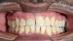
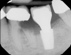
- Labialization of the dental implant, especially in the maxillary anterior region (figure 1)
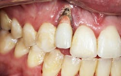
- Thinning of the hard and soft tissues around the dental implant (figure 2), which can increase the risk of a failed implant if tissue support is compromised
- Changes in occlusal patterns and force distribution, leading to possible fracturing of the implant components (figure 2a). Such changes can increase the risk of a failed implant and may require additional implant treatment to restore function and stability.
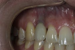
- Open contacts due to tooth migration, and cervical margin discrepancies in gingival height4 (figure 3)
Careful planning and ongoing monitoring are essential to ensure a successful procedure and minimize the risk of complications such as failed implants.
Patients at the highest risk for craniofacial growth that will impact dental implant positioning in future years fall into these risk factor categories:
- Female
- 35-years-old and younger
- Single-tooth replacements
- Replacing missing congenital teeth (especially maxillary incisors)
- Long, oval faces
- Thin gingival biotypes
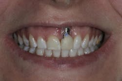
- High smile lines (figure 4)
Jaw development and gum recession around dental implants
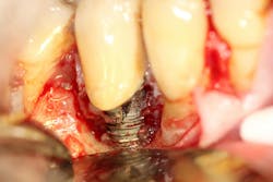
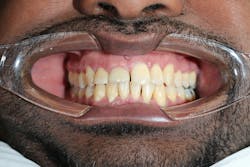
Gum recession around dental implants due to craniofacial development is a significant concern in implant dentistry, with the potential to undermine both the esthetic and functional success of dental implant procedures. When the gum tissue surrounding the implant site begins to recede, it can expose the underlying adjacent tooth root or the surface of the dental implant itself (figure 5). This not only affects the natural appearance of the artificial tooth but also increases the risk of dental implant failure and other complications.
Several risk factors other than jaw development contribute to gum recession around dental implants, including poor oral hygiene, gum disease (periodontal disease), teeth grinding, and even certain medical conditions. The esthetic impact of gum recession is particularly pronounced in the visible areas of the mouth, where the gumline plays a crucial role in the overall appearance of the smile. Receding gums can create an uneven gumline, expose the abutment screw or implant surface, and make the artificial tooth appear longer or mismatched compared to natural teeth. Additionally, exposed implant surfaces are more susceptible to bacterial accumulation, which can lead to peri-implantitis—a leading cause of dental implant complications and implant failure.
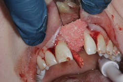
For patients who already exhibit signs of gum recession around their dental implants, advanced treatments such as bone grafting may be necessary to restore the supporting bone and stabilize the implant (figure 6). Addressing the underlying causes—such as correcting poor oral hygiene habits, managing teeth grinding, and treating existing gum disease—is critical to prevent recurrence and ensure the longevity of the implant.
Conclusion
In conclusion, it would be in the best interests of the treating dentist and the patient to explain that craniofacial growth may impact the need for reparations to the dental implant crown and/or the need for dental implant replacement prior to implant therapy. If a dental implant fails, the process of placing a new implant involves careful site preparation, consideration of bone healing, and timing to improve success rates.
In addition, patients who are at high risk may benefit from implant restorations that are retrievable (screw-retained), conventional fixed partial dentures, or removable partial dentures until that risk decreases. A risk assessment should be performed prior to dental implant therapy and informed consent obtained regarding the implications of craniofacial growth on future problems.
Frequently asked questions
Can dental implants move over time?
Dental implants themselves do not move once osseointegrated, but surrounding tissues and teeth can change.
Why do dental implants look worse years after placement?
Growth-related changes and gum recession can affect esthetics.
Who is at highest risk?
Younger patients, especially females with thin biotypes and high smile lines.
Can gum recession cause implant failure?
Yes, through peri-implantitis and bone loss.
How can complications be minimized?
Risk assessment, maintenance, and retrievable restorations.
Editor’s note: This article originally appeared in Perio-Implant Advisory, a chairside resource for dentists and hygienists that focuses on periodontal- and implant-related issues. Read more articles and subscribe to the newsletter.
Editor's note: Originally published September 13, 2016. Updated January 12, 2026.
References
- Testori T, Weinstein T, Scutellà F, Wang HL, Zucchelli G. Implant placement in the esthetic area: criteria for positioning single and multiple implants. Periodontol 2000. 2018;77(1):176-196. doi:10.1111/prd.12211
- Daftary F, Mahallati R, Bahat O, Sullivan RM. Lifelong craniofacial growth and the implications for osseointegrated implants. Int J Oral Maxillofac Implants. 2013;28(1):163-169. doi:10.11607/jomi.2827
- Oesterle LJ, Cronin RJ Jr. Adult growth, aging, and the single-tooth implant. Int J Oral Maxillofac Implants. 2000;15(2):252-260
- Varthis S, Tarnow DP, Randi A. Interproximal open contacts between implant restorations and adjacent teeth. Prevalence – causes – possible solutions. J Prosthodont. 2019;28(2):e806-e810. doi:10.1111/jopr.12980
About the Author

Scott Froum, DDS
Editorial Director
Scott Froum, DDS, a graduate of the State University of New York, Stony Brook School of Dental Medicine, is a periodontist in private practice at 1110 2nd Avenue, Suite 305, New York City, New York. He is the editorial director of Perio-Implant Advisory and serves on the editorial advisory board of Dental Economics. Dr. Froum, a diplomate of both the American Academy of Periodontology and the American Academy of Osseointegration, is in the fellowship program at the American Academy of Anti-aging Medicine, and is a volunteer professor in the postgraduate periodontal program at SUNY Stony Brook School of Dental Medicine. He is a trained naturopath and is the scientific director of Meraki Integrative Functional Wellness Center. Contact him through his website at drscottfroum.com or (212) 751-8530.