A quick technique for digital maxillofacial prosthetics
Editor's note: Originally published November 7, 2019. Reviewed May 24, 2022.
The Hippocratic oath dictates that clinicians do no harm and keep their patients’ best interests in mind at all times. Adhering to this doctrine means that we, as clinicians, must stay abreast of the latest treatment modalities, be as minimally invasive as possible, and always try to improve on treatment experiences and outcomes. Innovating and adapting digital workflows is part of this process given the enhanced patient experience and improved precision going digital provides.
Understanding the clear benefits that digital techniques offer for tooth- and implant-borne prosthetics, it seemed only natural to try to extend those benefits to maxillofacial prosthetics by adapting an off-label use of my TRIOS (3Shape) scanner. And so began my partnering with a local maxillofacial prosthodontist to facilitate the production of maxillofacial prosthetics by digitizing patients’ facial defects using my scanner.
Additional reading: The crown the lab made doesn't fit: Why does this happen?
In order to accurately use the TRIOS to complete a digital impression of a facial defect, one needs to thoroughly comprehend how the scanner builds a digital impression. Specifically, the TRIOS takes hundreds of images a second and stitches common landmarks within the images to successively build the impression. As a result, this scanner has a much easier time of scanning a surface with greater anatomical detail, while it is much more of a challenge to scan smooth surfaces.
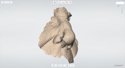
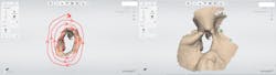
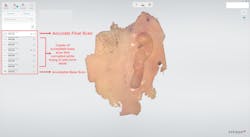
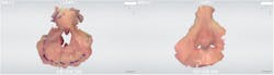
With this in mind, I recommend starting a maxillofacial prosthetic scan on the defect border in an area with the greatest amount of surface detail, and then following around the defect borders in a circular pattern working outward from the defect (figure 1). Due to the fact that smoother areas can confuse the software as to where it should be stitching in new images, it is recommended that if a scan seems complete but you want to continue trying to add to it, first copy it. If the software incorrectly stitches new images into the scan in a wrong location, you haven’t ruined an acceptable base scan (figure 2). This digital workflow works for all types of extraoral facial defects, from nasal to ocular to auricular (figures 3 and 4). In addition, it also allows us to scan family members for their intact facial features to be used as a design guide during the manufacture of the prosthetic (figure 5).
Some of the challenges with this off-label use of the TRIOS are due to current hardware and software limitations. The TRIOS tips are often too large to enter into smaller defects to completely capture sufficient border detail. Additionally, due to the nature of how a scan is built, it can be very challenging to capture the smooth, reflective scarred skin in burn victims and accurately build an impression of these tissues. These challenges aside, however, the benefits of this digital process in maxillofacial prosthetics is tremendous for greatly improving the treatment experience for a patient population that has often already suffered enough.


Author’s disclosure: Dr. Barbara Jurim is a paid consultant for 3Shape and receives compensation for lectures.
Editor’s note: This article originally appeared in Perio-Implant Advisory, a chairside resource for dentists and dental hygienists for issues relating to periodontal and implant medicine. Exclusive content from an academic perspective centers on complex care, solving clinical complications from a team-based approach through interdisciplinary management.

About the Author

Barbara Jurim, DDS
Barbara Jurim, DDS, is a prosthodontist who specializes in treating difficult restorative cases, implant dentistry, and challenging cosmetic cases. She graduated summa cum laude from Wellesley College with a biochemistry degree, and then attended New York University’s College of Dentistry (NYUCD) from which she graduated as valedictorian. She completed her studies in NYUCD's Postgraduate Prosthodontic Program. Dr. Jurim lectures extensively on the topics of cosmetics, full-mouth rehabilitations, and digital dentistry treatments. As the assistant director of digital dentistry at Touro College of Dental Medicine, she teaches digital treatment protocols. She is a founding member of the Jurim Dental Academy and TRIOSDoctors.com, where she provides her dental colleagues with advanced insight into CAD/CAM dentistry.