Diagnosing between ailing and failing implants: The haste to remove salvageable implants
The replacement of hopeless or missing teeth can be predictably achieved with endosseous dental implants (1) and has demonstrated high survival rates in the literature when restoring single teeth, multiple teeth, and/or completely edentulous cases. (2,3) Failures, however, do occur (4) and have been on the rise now that the term “implant success” has replaced the term “implant survival.” (5) Clinicians began to realize that an implant can still integrate or remain integrated showing no signs of mobility; however, a variety of other complications affecting the implant could still occur. Malpositioned implants, fractured implants or prostheses, and implants ailing with peri-implant mucositis or peri-implantitis are some of those complications that are seen in increasing numbers. (6) In addition, recent investigations now include the esthetic outcome of implant restorations, (7) and patient-based outcomes related to satisfaction (8) as inclusion criteria in their studies raise the bar on what is now considered implant success.
ALSO BY DR. SCOTT FROUM |Restoration of a damaged dental implant due to removal of a fractured screw: thinking outside the box
The term “ailing” implant has been described in the literature as an implant that has not failed, requiring explantation, but needs some form of therapy in order to preclude failure. (9) A biological complication of implant therapy characterized by inflammation in the soft tissues and progressive bone loss of supporting bone surrounding an osseointegrated implant is peri-implantitis, (10) and can be the cause of an implant status changing from healthy to ailing. Although the diagnosis of peri-implantitis has been defined and reviewed, proposed treatment has varied considerably. In fact, to date, there have been six systemic reviews regarding the treatment of peri-implantitis, (11) all using various treatment methods with different rationales for treating ailing implants vs. explantation. When deciding whether to treat an ailing implant as opposed to explanation, a similar decision matrix can be established as when deciding upon whether to save or extract a natural tooth. (12) For example, is the implant strategic in the alveolar arch; in other words, does it support a single-tooth restoration or a full prosthesis? If explanted, can the implant be replaced, or do anatomical and/or financial limitations exist? In addition, the age, medical history, smoking status, oral hygiene status, and — most importantly — desires of the patient all have to be taken into consideration.
ALSO BY DR. SCOTT FROUM |An alternative to conventional dental implants: short implants
One of the most important considerations when treating implants affected by peri-implantitis is the ability of the clinician to fully detoxify the implant surface during surgery. Surface decontamination using various techniques has been described in the literature (13) and is, in this author’s opinion, one of the most important considerations and keys to regenerative success around implants ailing from peri-implantitis. A more recent paper describing the use of a regenerative approach used to treat 51 peri-implantitis-affected implants with three- to seven-and-a-half-year follow-up reported encouraging results using a reproducible method, including a unique and meticulous method of thorough surface decontamination. (14) If the clinician is unable to thoroughly detoxify an implant surface, either a referral should be made or explantation of the implant is warranted since regenerative methods will have a much lower success rate. (15)
A recent review looked at the Er:YAG laser as a method of detoxifying implant surfaces. (16) This laser has the ability to effectively remove calculus and bacterial colonies from the impregnated implant surface without causing thermal damage to the implant and surrounding tissues. In addition, the ease of use and the focused beam allows access to often unattainable areas on the diseased implant, making a once recommended explanation into a regenerative possibility. The following case report describes the use of the Er:YAG laser for implant surface decontamination and concomitant regenerative therapy to treat an implant with advanced peri-implantitis.
A 52-year-old woman with a noncontributory medical history, taking no medications with no reported food or drug allergies, was referred to my office from a general dentist in order to obtain a second opinion. She had a lower right premolar implant placed in an oral surgery office five years ago that was suffering from severe peri-implantitis. (Fig. 1) Her first dentist told her that the implant had to be removed and that her treatment options were a fixed partial denture in the form of a three-unit bridge or a partial denture because re-implantation was not feasible. In addition, he sent her to a periodontal office that verified his statement, telling the patient that because of nerve proximity and the poor chance of ridge augmentation, re-implantation was unlikely. When she asked if saving the implant was possible, both the dentist and the periodontist told the patient that because of the level of bone loss, regenerative treatment would not work.
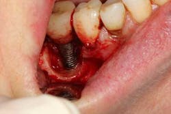
Fig. 4
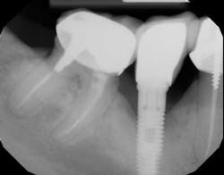
Fig. 5
Fig.6

References
1. Branemark PI, Hansson BO, Adell R, et al. Osseointegrated implants in the treatment of the edentulous jaw. Experience from a 10-year period. Scand J Plast Reconstr Surg 1977;16 (Supple): 1-132.
2. Den Hartog L, Slater JJ, Vissink A, Meijer HJ, Raghoebar GM. Treatment outcome of immediate, early, and conventional single-tooth implants in the aesthetic zone: A systematic review to survival, bone level, soft tissue, aesthetic, and patient satisfaction. Clin Periodontal 2008;35:1073-1086.
3. Adell R, Lekholm U, Rockler B, et al. A 15-year study of osseointegrated implants in the treatment of the edentulous jaw. Int J Oral Surg 1981; 10:387-416.
4. Koldsland OC, Scheie A, Aass AM. Prevalence of peri-implantitis related to severity of the disease with different degrees of bone loss. J Periodontol 2010;81:231-238.
5. Albrektsson T, Zarb G, Worthington P. The long-term efficacy of currently used dental implants. A review and prognosis criteria for success. Int J Oral Maxillofac Implants 1986;1:11.
6. Lang NP, Berglundh T. Peri-implant diseases: Where are we now? Consensus of the Seventh European Workshop on Periodontology. J Clin Periodontol 2011:38(Suppl. 11):178-181.
7. Furhauser R, Florescu D, Benesch T, Haas R, Mailath G, Watzek. Evaluation of soft tissue around single-tooth implant crowns: The pink esthetic score. Clin Oral Implant Res 2005;16:639-644.
8. Belser UC, Grutter L, Vailati F, Bornstein MM, Webre HP, Buser D. Outcome evaluation of early placed maxillary anterior single-tooth implants using objective esthetic criteria: A cross-sectional, retrospective study in 45 patients with a 2-4-year follow-up using pink and white esthetic score. J Perio 2009;80:140-151.
9. Peri-implant mucositis and peri-implantitis. A current understanding of their diagnosis and clinical implications. J Perio 2013;84:436-443.
10. Lindhe J, Meyle J. Peri-implant diseases: Consensus report of the Sixth European Workshop on Periodontology. J Clin Periodontol 2008;35(Suppl. 8):282-285.
11. Roos-Jansaker AM, Renvert S, Egelberg J. Treatment of peri-implant infections: A literature review. J Clin Periodontol 2003; 30:467-485.
12. McGuire, MK. Prognosis vs. actual outcome: A long-term survey of 100 treated periodontal patients under maintenance care. J Periodontol 1991;62:51-58.
13. Leonhardt A. Five-year clinical, microbiological, and radiological outcome following treatment of peri-implantitis in man. J Periodontol 2003;74:1415-1422.
14. Froum SJ, Froum SH, Rosen PS. Successful management of peri-implantitis with a regenerative approach: A consecutive series of 51 treated implants with 3- to 7.5-year follow-up. Int J Periodontics Restorative Dent 2012;32:11-20.
15. Gamal AY, Abdel-Ghaffar KA, Iacono VJ. A novel approach for enhanced nanoparticle-sized bone substitute adhesion to chemically treated peri-implantitis-affected implant surfaces: An in vitro proof-of-principle study. J Perio 2013;84:239-247.
16. Nevins M, Nevens ML, Yamamoto A, Yoshino T, Yoshino O, Wang C, Kim D. Use of Er:YAG laser to decontaminate dental implant surfaces in preparation for reestablishment of bone-to-implant contact. Int J Periodontics Restorative Dent. 2014;34:461-466.
About the Author

Scott Froum, DDS
Editorial Director
Scott Froum, DDS, a graduate of the State University of New York, Stony Brook School of Dental Medicine, is a periodontist in private practice at 1110 2nd Avenue, Suite 305, New York City, New York. He is the editorial director of Perio-Implant Advisory and serves on the editorial advisory board of Dental Economics. Dr. Froum, a diplomate of both the American Academy of Periodontology and the American Academy of Osseointegration, is in the fellowship program at the American Academy of Anti-aging Medicine, and is a volunteer professor in the postgraduate periodontal program at SUNY Stony Brook School of Dental Medicine. He is a trained naturopath and is the scientific director of Meraki Integrative Functional Wellness Center. Contact him through his website at drscottfroum.com or (212) 751-8530.