Partial Extraction Therapy (PET): Tooth-mediated ridge preservation
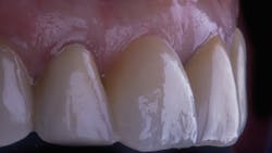
At the start of a new decade, we pause to reflect on the achievements in dental implant therapy that have made progress in our treatment of patients who have suffered tooth loss. Much has changed since the first reports of dental implants by Brånemark in Europe and Linkow in the US, both in 1969.1-2 Today, osseointegration is no longer an uncertainty, but rather, a highly predictable outcome.3 The success in terms of restoring a patient’s smile, however, is highly unpredictable.
Toward this, more than a decade ago, Fürhauser et al. already recognized the need for objective measures to evaluate our treatment success.4 These measures were updated in 2009 by Belser et al. to evaluate the optics of the restoration in addition to the soft tissue.5 The modified pink esthetic score discarded the evaluations for soft tissue texture and color. This underscored an era in implant dentistry that disapproved of immediate implant placement, in favor of early delayed treatment that would require the raising of flaps and augmentations after extraction and healing. These treatments typically result in less-than-desirable soft tissue changes.
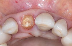
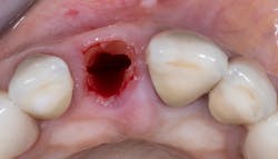
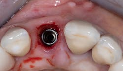
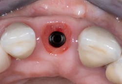
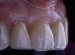
These concerns are with good reason. It is well understood that the loss of teeth results in the loss of the original periodontium.6 Hard tissue of the socket resorbs, then remodels. The overlying soft tissue architecture changes, typically in consequence to the hard tissue changes. The results are a healed site or sites that require significant effort on the clinician’s part to re-create the original anatomy—symmetrical gingival zeniths, full interproximal papillae, tissue of pleasing color, texture, and volume.
In the esthetic zone, these treatments are incredibly labor-intensive. They require considerable skill and knowledge of tissue biology, implant surgical technique, and prosthodontic tissue manipulation. In the event of an ideally esthetic outcome, the treatments are reparative regardless, correcting the missed opportunity to prevent tissue loss.
Today, the socket-shield technique is the subject of numerous case series and reports, four-year retrospective patient cohorts greater than 100 patients, human and animal histological research, randomized control trials to compare to conventional immediate implant placement, and so on.8-10 We have long passed the era of “Can this technique work?”
References
2. Linkow LI. The endosseous blade vent. Newsl Am Acad Implant Dent. 1969;18(2):15-24.
3. Buser D, Sennerby L, De Bruyn H. Modern implant dentistry based on osseointegration: 50 years of progress, current trends and open questions. Periodontol 2000. 2017;73(1):7-21. doi:10.1111/prd.12185
4. Fürhauser R, Florescu D, Benesch T, Haas R, Mailath G, Watzek G. Evaluation of soft tissue around single-tooth implant crowns: The pink esthetic score. Clin Oral Implants Res. 2005;16(6):639-644. doi:10.1111/j.1600-0501.2005.01193.x
5. Belser UC, Grütter L, Vailati F, Bornstein MM, Weber HP, Buser D. Outcome evaluation of early placed maxillary anterior single-tooth implants using objective esthetic criteria: A cross-sectional, retrospective study in 45 patients with a 2- to 4-year follow-up using pink and white esthetic scores. J Periodontol. 2009;80(1):140-51. doi:10.1902/jop.2009.080435
6. Chappuis V, Engel O, Reyes M, Shahim K, Nolte LP, Buser D. Ridge alterations post-extraction in the esthetic zone: A 3D analysis with CBCT. J Dent Res. 2013;92(suppl 12):195S-201S. doi:10.1177/0022034513506713
7. Hürzeler MB, Zuhr O, Schupbach P, Rebele SF, Emmanouilidis N, Fickl S. The socket-shield technique: A proof-of-principle report. J Clin Periodontol. 2010;37(9):855-862. doi:10.1111/j.1600-051X.2010.01595.x
8. Gluckman H, Salama M, Du Toit J. Partial Extraction Therapies (PET) part 1: Maintaining alveolar ridge contour at pontic and immediate implant sites. Int J Periodontics Restorative Dent. 2016;36(5):681-687. doi:10.11607/prd.2783
9. Gluckman H, Salama M, Du Toit J. Partial Extraction Therapies (PET) part 2: Procedures and technical aspects. Int J Periodontics Restorative Dent. 2017;37(3):377-385. doi:10.11607/prd.3111
10. Gluckman H, Salama M, Du Toit J. A retrospective evaluation of 128 socket-shield cases in the esthetic zone and posterior sites: Partial Extraction Therapy with up to 4 years follow-up. Clin Implant Dent Relat Res. 2018;20(2):122-129. doi:10.1111/cid.12554
Editor’s note: This article originally appeared in Perio-Implant Advisory, a newsletter for dentists and hygienists that focuses on periodontal- and implant-related issues. Perio-Implant Advisory is part of the Dental Economics and DentistryIQ network. To read more articles, visit perioimplantadvisory.com, or to subscribe, visit dentistryiq.com/subscribe.
About the Author
Howard Gluckman, PhD, MChD, BDS
Howard Gluckman, PhD, MChD, BDS, completed his dental training at the University of Witwatersrand in Johannesburg, South Africa, in 1990. After spending a number of years in general practice, he completed a four-year full-time degree in oral medicine and periodontics cum laude at Stellenbosch University in Cape Town, South Africa, in 1998. He was intimately involved in the development of the postgraduate diploma in implantology at both the Stellenbosch University and later at the University of the Western Cape. Dr. Gluckman recently completed his PhD titled, “Partial Extraction Therapy: Past, Present and Future,” summa cum laude at the University of Szeged in Hungary under the supervision of Professor Katalin Nagy. He is currently in full-time private practice in Cape Town, as well as the director of the Implant and Aesthetic Academy, a private postgraduate training facility that provides a complete postgraduate training program in implantology and esthetic dentistry in South Africa. The Academy is accredited by the State University of New York at Stony Brook and DentalXP. Dr. Gluckman has been involved in implantology training for more than 20 years.
