Dental case study: E.max Maryland bridges
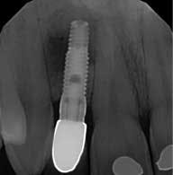
A 23-year-old patient presented with a healed bone level implant in site No. 10 to impress to fabricate the final restoration. The patient had a history of congenitally missing maxillary lateral incisors, and these missing teeth were restored with bone level implants post-orthodontic treatment in 2010. In 2013, a productive fistula was on the buccal tissue apical to the No. 10 implant. The implant had failed, so the surgeon replaced the failed implant and grafted the site.
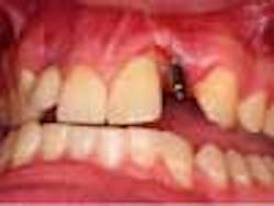
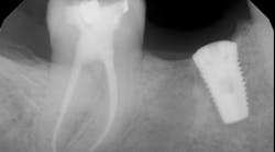
Upon examination, the No. 10 implant had a healing abutment placed and was noted to have circumferential loss of 3 mm to 4 mm of hard and soft tissue. This loss of tissue was significant enough that the collar of the implant fixture was now at the facial tissue margin. Also, tooth No. 9 had 5 mm of clinical attachment loss, and the tooth had been recently devitalized. A soft tissue lesion was also noted in the unattached tissue buccal to the apex of the No. 7 implant. The lesion appeared to be unproductive. A radiograph showed a large radiolucency around the apex of the No. 7 implant. The patient was referred to the periodontist to evaluate Nos. 9 and 10 for possible tissue graft and to find the origin of the lesion apical to the No. 7 implant.
The periodontist diagnosed the failed No. 7 implant with a large bone lesion around it, and he was not very confident in any significant gains of tissue in the No. 9 and 10 region. The patient at this point expressed a concern to have the definitive restorations placed without utilizing any implants, and for them to be as conservative as possible. To aid in the complexity, the patient had a limited time (10 days) to complete any treatment before he moved 2,000 miles away.
The patient was treatment planned alongside the periodontist to remove the No. 7 implant and graft the bone lesion. A cover screw was planned to be placed on the No. 10 implant and bury it under the soft tissue for esthetics. Two ceramic (e.max) Maryland bridges were placed to restore the missing laterals, bonding to both adjacent teeth. The possible complications were explained to the patient, and he consented to treatment. Initial data was gathered including photo grafts, mounted cast, and a diagnostic wax-up. The restorative plan was to be completed using a Planmeca E4D scanner and mill to fabricate the restorations.
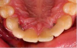
Three days before he left, the patient presented for treatment, the No. 7 implant had been removed, and the site had been grafted, along with No. 10 implant being submerged. A modified lingual veneer preparation was made on the adjacent teeth. The lingual prep served two purposes, the first to increase surface area for bonding and the second to increase the connector cross-sectional area. The ideal cross-sectional area is 16 mm² (*).
The preps were scanned, along with a bite registration, opposing dentition, and the diagnostic wax-up for cloning. The restorations were designed and milled from monolithic e.max HT blocks. The restorations were stained, glazed, and crystalized to final shade and luster.
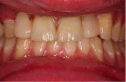
The restorations were etched with phosphoric acid, cleaned with Ivoclean (*), and bonded with universal bond (*). The preparations were isolated (isolation was very difficult in the area of No. 7 due to surgery the day before), etched, bonded, and the restorations were cemented. Special attention was paid to the occlusion on the FPDs, and all occlusal contact was removed from the connector area. Also, all anterior guidance was removed from the FPDs. The opposing dentition was reshaped to gain as ideal of a contact on the restorations as possible. The patient returned one prior to his leaving to verify his occlusion and post-treatment photographs. At this time, the patient was instructed about hygiene for the restoration and how to carefully function with the restorations. He seemed to be pleased with the functionality and esthetics of the restorations.
…
