Enhancing periodontal regeneration with the use of biologics
In the past decade, the regenerative potential of periodontal and furcation defects have become both more predictable and successful, largely due to the adjunctive use of biologic modifiers such as purified human platelet-derived growth factor-BB (PDGF-BB). (1-6) In the pivotal trial that lead to FDA approval of PDGF-BB for treatment in periodontal defects, the adjunctive use of PDGF-BB in combination with beta-tricalcium phosphate showed a greater potential of periodontal regeneration versus beta-tricalium phosphate alone.(3)Either beta-tricalcium phosphate or freeze-dried bone allograft (FDBA) reconstituted in PDGF-BB has shown a greater potential for periodontal regeneration in intrabony defects, furcation, and recession defects.(1,4,6,7,8) These results have been documented on clinical, histological, and microtomographic levels. (1,4,6,7,8)
ALSO BY DR. GREGORY PETTE |Vertical ridge augmentation: Using a titanium-reinforced high-density PFTE membrane
The advent of dental implants has dramatically changed the field of dentistry. However, we must not forget that many teeth with periodontal defects can be saved and treated predictably with proper diagnosis and treatment. Since dental implants are also prone to peri-implantitis and disease, we should not consider dental implants as a first choice in all cases of teeth with severe periodontal defects unless the teeth are deemed hopeless. It should always be our goal to maintain all teeth when possible so long as the results are going to be both predictable and cost-effective for the patient.
• Proper radiographic and clinical evaluation
• Testing tooth vitality, tooth mobility, and restorability; occlusal adjustments as needed
• Patient compliance
• Ideal and sound surgical techniques
• Proper case selection for regenerative procedures
• Proper communication between the patient, general dentist, periodontist, and dental hygienist
• The use of biologic modifier such as PDGF-BB as an adjunct to bone particulate and collagen membranes
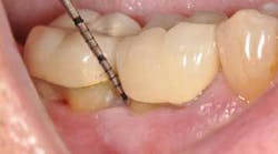
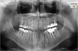
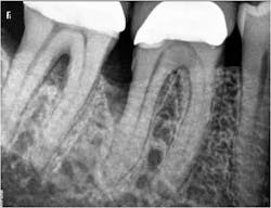
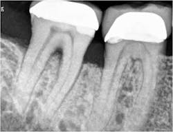
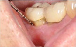
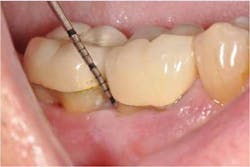
In the present case, a 61-year-old male was referred to my periodontal office by his general dentist for an evaluation of pain and bleeding to the lower right mandible. The patient’s medical history consisted of controlled type 2 (noninsulin dependent) diabetes and hypertension. Upon review of the panoramic radiograph (figure 1), the periodontal defects are not clearly evident, thus showing the importance of proper radiographs to detect periodontal defects. The periapical radiographs (figure 2) clearly show subgingival calculus, grade 1 furcation on No. 30, grade 3 furcation on No. 31, and an advanced intrabony defect on the distal of No. 30. Clinically, there was no mobility to either No. 30 or 31 and both teeth were vital. Tooth vitality is an extremely important diagnostic factor in treatment panning. Periodontal charting of the lower right was also performed, and the lower right exhibited bleeding and inflammation (figure 3).
The treatment plan for the lower right quadrant consisted of limited occlusal adjustments on Nos. 30 and 31 and osseous surgery of the lower right with regeneration using bone allograft (Straumann allograft, Straumann USA LLC, Andover, Massachusetts) and a resorbable collagen membrane (Optimatrix, Osteohealth, Shirley, New York) hydrated in PDGF (Gem21S, Osteohealth, Shirley, New York). The patient returned for surgery under conscious IV sedation (fentanyl, versed, dexamethasone, toradol, cleocin), and local anesthesia was established using lidocaine 1:100 epinephrine x 4 carpules.
Sulcular incisions were made from No. 27 to No. 31 on both the buccal and lingual in addition to a distal wedge. Full-thickness mucoperiosteal flaps were elevated, all soft tissue was removed, and roots were thoroughly planed and scaled using hand instruments and a piezo unit. Minor ostectomy, osteoplasty, and odontoplasty were performed in addition to thorough rotary curettage of the furcations on Nos. 30 and 31 and intrabony defect on No. 30. Thorough debridement of all soft tissue to sound bone in any periodontal defects is critical to obtaining ideal regenerative effects. The roots were treated with citric acid then thoroughly irrigated with sterile saline. The bone allograft and collagen membrane were both hydrated in PDGF-BB for 20 minutes, then the allograft was placed in the intrabony and furcation defects then covered with the collagen membrane; the flaps were then approximated using 4-0 chromic suture.
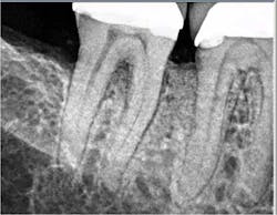
A postop radiograph was taken (figure 4) and the patient was recovered and dismissed with an escort. The patient was placed on postoperative medications (doxycycline 100 mg for 10 days, Motrin 800 mg, and narcotic pain management as necessary), in addition to oral hygiene instructions including the use of locally applied chlorhexidine twice daily.
At the two-week postop appointment, the patient was doing well and started to resume normal hygiene. Care was taken to ensure that at any future dental appointments for the following six to nine months no dental professional periodontal probes or scalers were inserted subgingivally into the regenerative sites, which would compromise the results.
The patient returned for a postop appointment at 5.5 months and a new radiograph (figure 5) and clinical photographs (figures 6 and 7) were taken. Excellent regeneration is evident at both sites radiographically and clinically. New periodontal charting was done (figure 8) showing near-complete regeneration of the intrabony defect on the distal of No. 30 and a great improvement in the furcation of No. 31. There is no bleeding or inflammation present at this time.
ADDITIONAL READING …
Periodontal regeneration: Back to the future
Manage, repair, or regenerate periodontal disease?
References
1. Camelo M, Nevins ML, Schenk RK, Lynch SE, Nevins M. Periodontal regeneration in human Class II furcations using purified recombinant human platelet-derived growth factor-BB (rhPDGF-BB) with bone allograft. Int J Perio and Rest Dent. Jun. 2003;23(3):213-225.
2. Nevins M et al. Periodontal regeneration in humans using recombinant human platelet-derived growth factor-BB (rhPDGF-BB) and allogenic bone. J Periodontol. Sept. 2003;74(9):1282-1292.
3. Nevins et al. Platelet-derived growth factor stimulates bone fill and rate of attachment level gain: Results of a large multicenter randomized controlled trial. J Periodontol. Dec. 2005;76(12):2205-2215.
4. Nevins M, Hanratty J, Lynch SE. Clinical results using recombinant human platelet-derived growth factor and mineralized freeze-dried bone allograft in periodontal defects. Int J Perio Rest Dent Oct. 2007;27(5):421-427.
5. Nevins M et al. Platelet-derived growth factor promotes periodontal regeneration in localized defects: 36-month extension results from a randomized controlled, double-masked clinical trial. J Periodontol. 2013;84:456-464.
6. Reynolds M et al. Periodontal regeneration—Intrabony defects: A consensus report from the AAP regeneration workshop. J Periodontol 2015;86(suppl):S105-107.
7. Ganeles J, Pette GA. Regenerative treatment for recession defects using purified human platelet-derived growth factor-BB, particulate grafts and coronally repositioned flaps. Clin Adv in Periodontics. May 2012; 2(2):57-64.
8. McGuire MK, Scheyer T, Nevins M, Schupbach P. Evaluation of human recession defects treated with coronally advanced flaps and either purified recombinant human platelet-derived growth factor-BB with beta-tricalcium phosphate or connective tissue: A histologic and microcomputed tomographic examination. Int J Perio Rest Dent. Feb. 2009;29(1):7-21.
