Importance of soft tissue in interdisciplinary cases: A case study involving orthodontics, periodontics, dental implants
The importance of keratinized soft tissue around both dental implants and natural teeth has been demonstrated in the literature to support effective home care, prevent sensitivity and tissue breakdown, as well as aid in esthetic value. The etiology of soft-tissue loss is multifactorial and can include improper home-care methods, genetically thin tissue, trauma to the area, fenestrations in the bone, and/or orthodontic movement. Unfortunately, clinical exams performed by clinicians can often overlook the need for soft-tissue enhancement until the patient becomes symptomatic and/or desires esthetic improvement. In addition, soft-tissue augmentation prior to orthodontic treatment, prosthodontic treatment, and implant therapy can be of tremendous benefit to ensure a long-term, successful result. This case study highlights the use of soft-tissue augmentation prior to orthodontic and implant therapy to enhance the zone of keratinized tissue, increase root coverage, and improve esthetics.
ADDITIONAL READING |Interdisciplinary management of a complex maxillary implant restoration
Case example
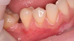
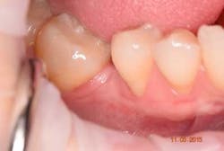
This 35-year-old female with a noncontributory medical history presented to private practice with a chief complaint of “a possible crack on the upper right canine and generalized teeth sensitivity when brushing.” She was missing the upper left and lower right first molar. Tooth No. 30 was lost at the age of 9, and the second molar, tooth No. 31, was mesially inclined and drifted into the area of No. 30. Both horizontal and vertical ridge deficiency was noted, and inadequate space for an implant was present. Her lower right quadrant demonstrated Class I and II Miller recessions with less than 2 mm of keratinized tissue (figure 1).
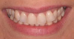
Because of her high-stress job and infrequent use of her night-guard, clenching and bruxism habits were reflected in her dentition. In addition to malocclusion and crowding, her upper right canine has a lingual enamel chip with an incipient crack line (figures 2a and 2b).
In order to correct the patient’s restorative and orthodontic concerns, as well as her chief complaint of sensitivity, an interdisciplinary treatment plan was presented and agreed upon in the following sequence:
- Fluoride varnish, improved use of night-guard, proper home-care instruction
- Free gingival graft to improve zone of keratinized tissue and prophylactically prevent further recession
- Orthodontic correction to improve crowding, occlusion, spacing, and upright No. 31 for future restorative and implant dentistry
- Ridge augmentation with hard and soft tissue to improve vertical and horizontal ridge defects
- Implant placement for Nos. 14 and 30
- Prosthetic treatment
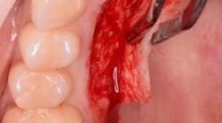
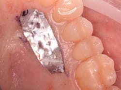
The following will now describe the free gingival autograft surgery that was used to augment the zone of keratinized tissue prior to orthodontic therapy. Three carpules of 3% Septocaine (Septodont, 1.7 ml carpule) was used as a local anesthetic for both infiltration of the donor palate area and inferior alveolar/mental nerve block of the recipient site. Injecting local anesthetic in the mucosa of the recipient site helps to identify the MGJ (mucogingival junction). A submarginal, split-thickness incision with a 15 blade was used to prepare the recipient site. Using aluminum foil to measure the amount of graft needed (figure 3), a full-thickness, free gingival graft was harvested from the upper right palate in the area of Nos. 3-6. Careful attention is paid not to harvest the site too anteriorly to avoid ruggae as well as limit increased post-operative pain.
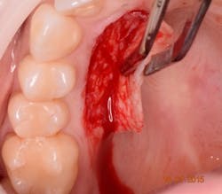
The harvesting technique (figure 4) should aim for a graft thickness between 1-3 mm and be 15-25% larger than the desired final size. Harvesting a larger amount of graft is warranted due to both initial (immediate) and secondary contraction of the graft during healing. A thin graft will have less primary and more secondary contraction, where as a thick gingival graft will have more primary and less secondary contraction. In general, a harvested soft-tissue graft can be expected to shrink on average of 15% to 20%.
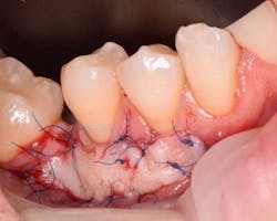
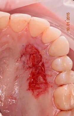
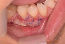
The free gingival graft was secured using 6.0 Prolene sutures in single interrupted fashion (figure 5). The gingival margin of the vestibular mucosa was fixed to the periosteum using the same suture. The donor site was sutured with 4.0 chromic gut resorbable sutures in single interrupted fashion (figure 6).
Plaque control after surgery was maintained using a very soft brush, avoiding the surgical side for one week and also using a PerioSciences antioxidant gel. No periodontal dressing was used. Healing was uneventful, and the patient did not have discomfort after one week post-operatively (figure 7).
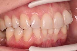
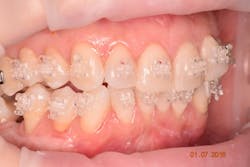
The patient demonstrated excellent healing one month post-operatively (figure 8) with a significant increase in keratinized tissue. This increase in soft tissue will aid in the prevention of further recession during orthodontic therapy (figure 9) as well as assist in the coronal advancement of tissue post orthodontic therapy for the purposes of root coverage. Most importantly, the patient was extremely happy with the result.
ADDITIONAL READING |A unique method of debriding a full-arch, maxillary prosthesis supported by dental implants
