How to increase safety and reduce complications during sinus lift

March 13, 2013
Maxillary sinus augmentation (or sinus lift) is an effective surgical procedure used to increase bone in the posterior maxilla and allow placement of dental implants. Alessandro Geminiani, DDS, MS, describes a recently developed technique that significantly reduces the incidence of complications associated with this procedure.

Introduction

Maxillary sinus augmentation (or sinus lift) is a very effective surgical procedure used to increase bone in the posterior maxilla and, therefore, allow placement of dental implants. The complications most frequently encountered during this type of surgery are laceration of an intraosseous artery (causing bleeding) and perforation of the Schneiderian membrane (the membrane lining the maxillary sinus cavity). In case of bleeding, the procedure has to be suspended (sometimes aborted) until the bleeding is controlled. In case of perforation of the Schneiderian membrane, a repair must be attempted, increasing considerably the length of the procedure and consequently the postoperative discomfort and incidence of complications. The conventional technique for maxillary sinus augmentation involves the use of a high-speed handpiece for the removal of the thin cortical plate that constitutes the lateral wall of the maxillary sinus. In the vast majority of the cases, this bony wall is less than 1 mm (Neiva et al 2004). With the conventional technique, perforation of the Schneiderian membrane occurs in approximately 40% of the sinus lift procedures. A recently developed technique (Geminiani et al 2011) significantly reduces the incidence of this type of complications (Geminiani et al, in press).

RELATED |And you thought dental plaque was bad: preventing evolution of biofilms

RELATED |A review of the maxillary sinus

Description of the technique

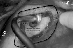
1. A full-thickness flap is reflected to gain access to the lateral wall of the maxillary sinus (Fig. 1). In this area the bone is normally very thin, usually less than 1 mm.
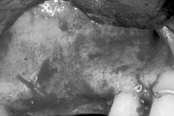
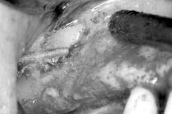
Fig. 12. The sonic handpiece (Fig. 2) is the ideal device for the preparation of the lateral window. The diamond-coated insert does not rotate, but vibrates (at 6 kHz). This allows a precise and safe osteotomy, reducing significantly the incidence of perforations (Weitz et al, in press).
Fig. 23. The osteotomy of the inferior border is completed. The Schneiderian membrane has a grayish halo, making it easier to distinguish from the adjacent tissues. The cavitation effect of the sonic instrument produces temporary hemostasis, improving the visibility of the surgical field. Once the inferior border is completed, we can proceed to the preparation of the mesial, superior, and distal border (Fig. 3).

Fig. 3
4. The sinus membrane is separated from the bony wall. Perforations of the membrane occur frequently during this phase. To increase safety and reduce complications, the air-driven sonic handpiece is used, coupled with the discoid insert. The insert is activated (vibrating and irrigated by the water spray) and then placed between the lateral bone wall and the Schneiderian membrane (Fig. 4).
Fig. 45. The sinus is grafted with the material of choice. Once the graft has been placed in the subantral cavity, the lateral window osteotomy is covered with a collagen membrane. The illustration clearly shows the criteria that I consider important in the placement of the absorbable membrane (Fig. 5).
Fig. 56. Primary closure is fundamental for the success of this procedure (Fig. 6). If primary closure cannot be achieved by flap replacement, a periosteal releasing incision is indicated. More details on how to perform a safe and effective periosteal releasing incision can be found in an excellent article recently published (Romanos 2010).
Fig. 6
Dr. Alessandro Geminiani received his DDS degree from the University of Siena (Italy) in 2004, summa cum laude. He then pursued a Master of Science in fixed prosthodontics and prosthetic dental materials, graduating in 2006 with highest honors. He continued his education in the USA by pursuing a two-year certificate in advanced general dentistry (2009), a certificate in periodontology (2012), and a Master of Science in clinical investigation (2012). Dr Geminiani is fluent in English, Spanish (Castellano), and Italian. You can find his full CV on LinkedIn. Dr. Geminiani lives in Houston, Texas. His practice focuses on bone grafts, gingival grafts, periodontal surgery, and dental implants. Visit his blog about periodontics and dental implants at LovePerio.com.