Computer-guided dental implant surgery: evolving, efficient, esthetic
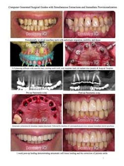
By Richard Nejat, DDSIn 1987 computed tomography (CT) was introduced into dentistry to add another dimension to dental implant treatment planning. This technology allowed clinicians for the first time to evaluate anatomic structures with a higher level of accuracy.In 1999 dental implant planning applications were developed, allowing interactive planning of virtual implants in 2-D and 3-D. The use of radiopaque templates/scanning appliances at the time of the CT scan made it possible for the prosthetic outcome to be incorporated into interactive presurgical planning. This advancement paved the way for an association between radiographic anatomic interpretation, prosthetic treatment planning, and precise surgical execution. Through the use of stereolithography and CAD/CAM technology, surgical templates could be fabricated to help clinicians place implants in a well-planned preoperative/prosthetic manner, rather than intraoperative planning, which is often surgical-driven.The use of surgical templates can benefit the patient as well as the dental team (restorative dentist, surgical specialist, and laboratory technician). The work performed can be more accurate and less invasive than in traditional cases. The ability to transfer the desired three-dimensional position of implants from the virtual model to the mouth has made this a more efficient outcome. The surgical template essentially has two functions: one for the surgical phase and one for the laboratory phase. It is used as a laboratory template to fabricate a master model, which is used to create the premanufactured implant-supported prosthesis.Following is a case example of the benefits of CAD/CAM computer-generated surgical guides. This case involves simultaneous full-mouth extractions, incisionless computer-guided implant placement, and immediate insertion of a premanufactured cast metal, reinforced, screw-retained 12-unit bridge. Immediately loaded prosthetic restorations have been shown to help support the patient's gingival architecture and allow him or her to function with an esthetic restoration while healing.
Click here to see the photos larger.Author bioDr. Richard Nejat is board certified by the American Board of Periodontology. His practice is on the forefront of computer guided implant dentistry and minimally invasive dental surgery. He is a course instructor in various continuing educational seminars, symposiums to colleges, and dental societies. Dr. Nejat attended Drew University and continued his education at New York University, where he attained his doctorate of dental surgery. Dr. Nejat was elected to membership in Omicron Kappa Upsilon, the National Dental Honor Society recognizing academic and clinical excellence in dentistry. He earned his certificate in periodontics from the State University of New York at Stony Brook. Dr Nejat is currently clinical assistant professor in the department of implant dentistry and periodontics at New York University and maintains private practices in Manhattan and Nutley, N.J.

