Interdisciplinary management of a complex maxillary implant restoration

Oct. 21, 2014
Although implants enjoy a high success rate in the literature, the restorative challenge continues to be achieving a desirable result in the maxillary anterior segment. This challenge is further exacerbated by patients presenting with malocclusion, soft tissue loss, hard tissue loss, a high lip line, and/or high esthetic demands. A patient who exhibits all of these factors can only be managed through proper treatment planning. The following case discussion from Drs. Peter Mann and Scott Froum highlights the importance of proper treatment planning and communications that take place between the implant surgeon and restorative dentist.

Although implants enjoy a high success rate in the literature, the restorative challenge continues to be achieving a desirable result in the maxillary anterior segment. This challenge is further exacerbated by patients presenting with malocclusion, soft tissue loss, hard tissue loss, a high lip line, and/or high esthetic demands. A patient who exhibits all of these factors can only be managed through proper treatment planning. The following case discussion highlights the importance of proper treatment planning and communications that take place between the implant surgeon and restorative dentist.

ALSO BY DR. PETER MANN | Narrow diameter dental implants in the esthetic zone

ALSO BY DR. SCOTT FROUM | Restoration of a damaged dental implant due to removal of a fractured screw: thinking outside the box

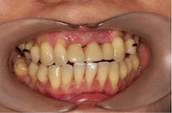
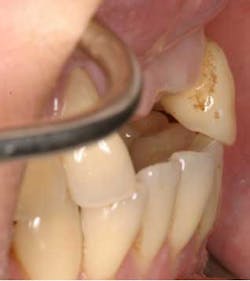
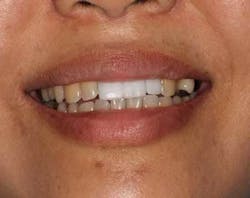
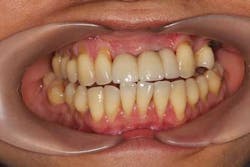
A 43-year-old female with a noncontributory medical history taking no medication and having no known food/drug allergies presented to my general dental office with a chief complaint of having “loose teeth with an underbite.” Clinically, she had generalized, moderate, chronic periodontal disease and was missing teeth Nos. 8 through 10. She had a Class III malocclusion with pathologic flaring of her remaining anterior teeth. She had both vertical and horizontal tissue loss in the No. 8 through 10 region. The patient wore a transitional partial denture to replace the anterior missing teeth that was placed in edge-to-edge occlusion by her previous dentist. (Fig. 1) She did not like having a removable prosthetic and desired a fixed option in the form of implants to replace her missing teeth. I sent her to the periodontist for a periodontal consult for her remaining dentition and dental implants to replace her missing front teeth.

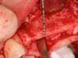
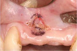
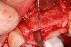
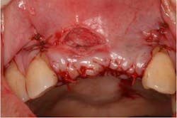
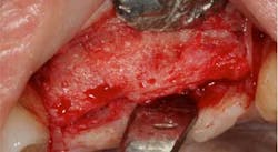
Fig. 1From a surgical perspective, after the patient presented for consult, diagnostic models and a cone-beam scan were taken. From the CT scan and clinical photos, a severe horizontal bony defect was noted requiring ridge augmentation procedures prior to implant therapy. (Figs. 2 and 3) In addition, because of the moderate periodontal disease, the patient was going to need periodontal therapy prior to implant therapy in order to decrease the bioburden. These findings were discussed with the restorative dentist and patient. After treatment was accepted, the patient was put through initial and surgical periodontal therapy. Once her hygiene was optimal, a surgical guide was fabricated in order to facilitate her contour augmentation. On the day of her augmentation surgery, a laser frenectomy was first performed to free up her tissue for advancement. (Fig. 4) A split-thickness flap was elevated, leaving a periosteal tissue layer (Fig, 5), which was later elevated. (Fig. 6) This method allows both stabilization of the membrane and easy advancement of the flap.

Fig. 2
Fig. 3
Fig. 4
Fig. 5
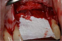
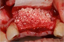
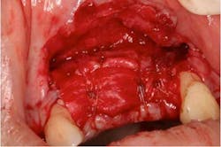
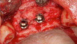
Fig. 6After full-ridge exposure was achieved and a 2 mm crestal ridge was noted (Fig. 7), a combination of mineralized corticocancellous allograft and autogenous bone graft was used with a collagen membrane. (Figs. 8 and 9) The membrane was sutured to ensure fixation. (Fig. 10) The flap was coronally advanced, achieving tension-free primary closure. (Fig. 11) The ridge was augmented according to where the surgical guide indicated the future position of the teeth. Six months later, a full-thickness mucoperiosteal flap was made and the area was reentered for implantation. The regenerated ridge now measured 8 mm in width (Fig. 12) allowing for two, 4 mm platform implants to be placed in the No. 8 and 9 region. (Fig. 13) The area was allowed to heal for an additional six months before Stage II was performed.

Fig. 7
Fig. 8
Fig. 9
Fig. 10

Fig. 11
Fig. 12
Fig. 13

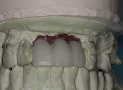
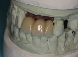
From a restorative perspective, now that the implants were placed in a restoratively driven manner, the main considerations were to address the patient’s malocclusion and correct her edge-to-edge bite while delivering optimal esthetics. In order to accomplish this goal, a framework was fabricated with a wax-up on the casts. (Fig. 14) By utilizing this method at the framework try-in visit, we were able to give the patient an extremely accurate preview of what her final prosthetic would look like.

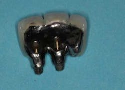
Fig. 14The patient was satisfied with her overbite, overjet, and overall appearance of the three-unit implant bridge. (Fig. 15). Our final concern was the risk of porcelain fracture due to the patient’s deep bite. She would occlude on the lingual of her new implant bridge with more force than someone with Class I occlusion. By making this prosthesis screw-retained, we will be able to retrieve this bridge in case of future porcelain fracture without destroying the framework. (Fig. 16) We can either repair the porcelain fracture or completely remake the porcelain on top of the existing framework. The lingual, where the patient is occluding, was also left in metal to further reduce the risk of possible porcelain fracture.

Fig. 15
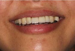
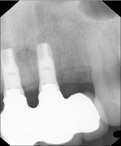
Fig. 16After all the concerns and risks were addressed, the case was sent to the lab for finalization. (Fig. 17) Six months post final insert, the patient is happy with her esthetics and function, (Figs. 18 and 18a) and her radiographs demonstrate success. (Fig. 19)

Fig. 17
Fig. 18
Fig. 18a
Fig. 19
Peter Mann, DDS, FICOI, FAGD, graduated from the New York University College of Dentistry in 2008, and completed his General Practice Residency at Kings County Hospital in 2009. He is the director of Smile Arts of NY, a comprehensive general practice in midtown Manhattan with an emphasis on cosmetic and implant dentistry.
Scott Froum, DDS, is a periodontist and co-editor of Surgical-Restorative Resource e-newsletter, as well as a contributing author for DentistryIQ and Dental Economics. He is a clinical associate professor at the New York University Dental School in the Department of Periodontology and Implantology. Dr. Froum is in private practice in New York City. You may contact him through his website at www.drscottfroum.com.