Have infection, will travel: Paving the path to dental implant failure
Why did this dental implant fail? You did everything right during your implant treatment. You properly diagnosed and treatment planned the patient’s implant therapy. During surgery, you (or the implant surgeon) placed the implant in a prosthetically driven orientation. You (or the restorative dentist) placed the abutment and crown on the dental implant with the correct contours, perfect fit, and (if not screw retained) without excess cement. The patient adhered to the follow-up appointments and had good oral hygiene. The follow-up visits showed good bone levels around the implant . . . and then one day there was implant infection and loss of integration. Why?
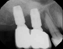
This is a scenario that has happened in practice and, yet, looking back at the radiographs and clinical pictures, there was one big oversight. The adjacent tooth next to the implant had pathology. As clinicians, it can be easy to focus on the area of treatment. When examining post-implant placement radiographs, bone levels are often the first thing we examine. After restoration, we look at excess cement, abutment fit, crown fit, and bone levels. All of these factors are important, but just as important is to examine the status of the adjacent tooth (or implant) and check for periapical radiolucencies and/or dental caries.
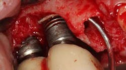
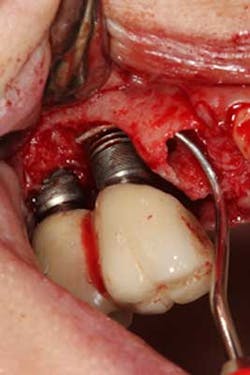
There have been numerous case reports in the literature demonstrating infections of dental implants caused by pathology from the adjacent natural tooth. (1) These infections from adjacent teeth can travel quite quickly to the proximal dental implant (figures 1a and 1b) and cause a rapid degree of bone loss. Treatment can consist of loss of teeth, loss of the dental implants, and/or costly regenerative repair (figure 2). (2) This situation is highly preventable if detected early and endodontic therapy is initiated.
The next time you take a post-placement radiograph, look at the adjacent tooth—especially the apex.
MORE CLINICAL TIPS FROM DR. SCOTT FROUM . . .
References
1. Tseng CC, Chen YH, Pang IC, Weber HP. Peri-implant pathology caused by periapical lesion of an adjacent natural tooth: a case report. Int J Oral Maxillofac Implants. 2005;20(4):632-5.
2. Tözüm TF, Sençimen M, Ortakoğlu K, Ozdemir A, Aydin OC, Keleş M. Diagnosis and treatment of a large periapical implant lesion associated with adjacent natural tooth: a case report [published online May 3, 2006]. Oral Surg Oral Med Oral Pathol Oral Radiol Endod. 2006;101(6):e132-8. Accessed April 10, 2016.