Are you sure I have a cavity?
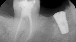
This was the first question out of my patient’s mouth when I diagnosed him with a cavity at his new patient exam. Of course he had to ask … his last filling was when he was 14 (he is now 37), and understandably there was an underlying tone of doubt in his voice. After all, what dentist in his or her right mind would look at tooth No. 29 and say, “That’s a cavity”? (Figs. 1 and 2)
Fig. 1
In dental school, we were taught to rely on a sharp explorer and sharp eyes to diagnose caries. Upon initial examination, I did not consider this to be a cavity. If we can’t see it, it’s not there, right? Why doubt what we were taught? The answer is simple. Because we can’t SEE everything that needs to be diagnosed, we therefore need to utilize adjunct diagnostic tools to be sure we are thorough in our examinations.
ALSO BY DR. STACEY SIMMONS |Head, neck, and oral cancer: A general reference for treatment
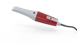
The DIAGNOdent (by KaVo) is one of several types of caries detecting adjuncts that can be a part of every practicing dentist’s arsenal of gadgets — and I know we have plenty of them. The hidden caries phenomenon is something we all face daily — is it there or isn’t it? Is it stain or an actual cavity? Should I open it up or just “watch” it? Should I be conservative or aggressive in my treatment approach?
The DIAGNOdent permits us to DIAGNOSE infection that we otherwise miss and therefore treat. Having such a remarkable tool that apportions us to painlessly, easily, and quickly assess a potential caries situation where there would otherwise be doubt is invaluable. Its readings, when used correctly, are reproducible, accurate, and furthermore promote confidence in the patient/doctor relationship. This is an investment that easily pays for itself time and time again.
How does the DIAGNOdent work? When a tooth presents with altered surfaces, it will fluoresce when exposed to a certain wavelength of light. The DIAGNOdent operates at a wavelength of 655nm. (1) When applied to a healthy tooth surface, little to no fluorescence is present and thus a low numerical reading is displayed. The more compromised the tooth structure, the higher the reading. Readings range from 0 to 99 — the size of caries or demineralized tooth structure is directly proportionate to the reading.
In the aforementioned case, the reading on the occlusal surface was a 46 (see Figs. 3 through 8 below). Look at what was underneath. Surprised? I often wonder how many times per day lesions like this are going undiagnosed.
After the final smear layer was removed, the tooth was filled with Grandio resin. Afterwards, I showed the photographs to my patient. Any doubt that he had was immediately erased. Most importantly, he was grateful and his trust in me was solidified.
Figs. 3 through 8 below ....References


