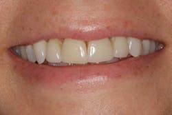
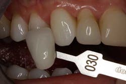
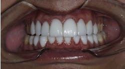
A 35-year-old woman presented with discoloring of the central and lateral incisors, as well as gingival recession around the same teeth (Figs. 1 and 2). The patient had veneers placed approximately 15 years prior. The overall shape and length were acceptable to the patient, but she wanted to improve the color and appearance of the discolored margins. Her natural canines were an O2O shade (Fig. 3), but the central and lateral incisors were much darker. A full series of photographs and records were obtained. The patient decided to bleach her teeth prior to shade selection for her new veneers.
Fig. 1: Preoperative full-face view helps harmonize dental esthetics with facial esthetics.
Fig. 2: Preoperative photograph indicates the relation of the teeth to the lip line.
Fig. 3: Shade tabs were previewed.
Material selection
Pressable lithium disilicate (IPS e.max Press, Ivoclar Vivadent, Amherst, N.Y.) was the material of choice. Ideal for esthetically challenging cases, lithium disilicate demonstrates lifelike translucency due to a relatively lower refractive index and superior optical properties, compared to traditional all-ceramic materials. A variety of translucencies and opacities — including high translucency (HT, 16 A-D, and 4 BL Shades), low translucency (LT), medium opacity (MO), and high opacity (HO) — is available. Different brightness effects can be reproduced with the availability of three brightness values and two opalescent shades (Value [V], Opal [O]). Providing minimally invasive preparation for treatment that is gentle to the tooth structure, lithium disilicate ingots offer versatile use and a comprehensive range of indications. Press technology guarantees a high accuracy of fit and natural-looking shades, and optimal light transmission provides highly esthetic, lifelike restorations.
RELATED |Avoiding complications bonding lithium disilicate
Clinical preparation protocol
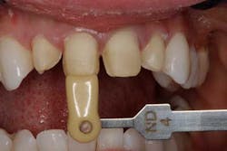
The patient was anesthetized and the old porcelain removed. Preparation was carried through the contact, slightly subgingival to allow proper interproximal contours. A final impression was taken. Preparation photographs with shade tabs were obtained (Fig. 4), as well as a stick bite and bite registration. Bisacryl provisionals were fabricated from a prepreparation model, making only slight modifications to improve esthetics.
Fig. 4: Preparation shade was determined as ND4.
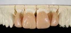
The provisionals were evaluated four days later (Fig. 5), at which time the patient was happy with the color and esthetics. Occlusal stops remained in the enamel, therefore only incisal edge position needed to be evaluated for esthetics, speech, and function. Once confirmed, a series of photographs and a stone model of the provisionals, as a starting point for shape and form, were taken and forwarded to the laboratory to duplicate the incisal edge position.
Fig. 5: Photographs of the provisionals were taken and a stone model created.
Laboratory protocol
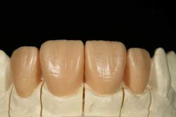
A master model was created, and a Sil-tech matrix (Ivoclar Vivadent, Amherst, N.Y.) of the provisional models was placed over the master model (Fig. 6). Data from the provisionals, complete with incisal edge and form, was transferred to the master model using wax injection (Fig. 7). Wax was then added to the master model and shaped. The incisal edge was lengthened and the midline embrasure made vertical. The transition from embrasure to facial surface was structured and the mesial lobe shaped to create the planned esthetic result.
Fig. 6: A sectioned working model was created.
Fig. 7: Using wax injection, data from the provisional stone model was transferred to the working model.
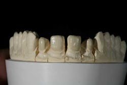
Surface morphology was addressed by tucking in the distal, and because the teeth seemed slightly oversized, the desirable amount of reduction was performed. Referencing the incisal silhouette and midline embrasure, lobe formation shapes were refined, narrowing the appearance of the distal lobe by creating more deflective surface and defining the fine surface anatomy, creating triangular depressions (Fig. 8). Due to its hardness, it is more efficient to perform surface texture and detailed waxing — two crucial steps, especially when using a monolithic lithium disilicate material — than to grind the restorations.
Fig. 8: An enhanced waxup indicates contour refinements and detailed surface texture.
The restorations were sprewed and invested in the traditional manner for pressed ceramics, and a fast burnout technique was administered. After they were pressed using the IPS e.max Press Opal 1 lithium disilicate ingot, the restorations were divested and the sprews ground down using a lithium disilicate and zirconia grinder (Brasseler, Savannah, Ga.). Next, an 863 carbide bur (Brasseler, Savannah, Ga.) was utilized to create surface morphology between the teeth, delineating the tooth as individual in relation to the adjacent tooth. Positioning the contact and controlling the light into the embrasure form is critical to achieve superior esthetics. A medium grit, lithium disilicate grinder (Brasseller, Savannah, Ga.) was used to clean up the lingual area close to the margins, making it possible to get right up against the margin without chipping.
At this time the restorations were ready for staining. The surface was dampened with a small amount of stain liquid. It is important that stain liquid be applied underneath and between the restoration and the composite die. This step determinedly affects the amount of stump shade show-through and plays an important role in attaining the natural color of the dentition. For instance, 1.2 mm of Opal 1 will have quite a different affect than .4 mm facial thickness. Viewed side by side, the thicker appear brighter than the thinner restorations. In this case, the thinner restorations are a BL4, while the thicker present as a BL1 shade.
At this point, stain was applied from gingival to incisal to achieve a color gradient effect. Stain was blended to achieve natural color gradation and applied to all restorations in this manner. Although the Opal ingot is already translucent, a small amount of incisal blue was added for enhancement. After the stains were fired, a glaze layer was applied. Finally, a small amount of white stain was added to the glaze to attain a white halo effect. Although requiring a fairly thick restoration, simply by choosing the appropriate ingot, an Opal 1 in this case, the optical qualities of the natural tooth enamel were well matched, eliminating the need for cutback and layering.
Final seating
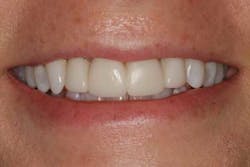
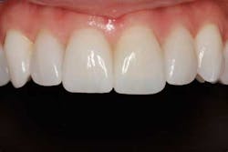
After the patient was anesthetized, the provisionals were removed by sectioning with a very thin carbide bur at high speed, but with very light pressure. The preparations were cleaned, and each unit was tried-in separately for marginal fit. Next, all four veneers were tried-in together with a small amount of transparent try-in paste to confirm contacts and esthetics. The porcelain was prepared and the teeth treated with a fifth generation bonding agent (Excite F single), before seating with a light-cure-only veneer resin cement (Variolink Veneer). Occlusion was refined and the margins polished. Final presentation (Fig. 9)Fig. 9: Final presentation
Conclusion
Lithium disilicate restorations (IPS e.max Press) enable dentists to offer patients a conservative alternative while transforming the appearance of their smiles. With lithium disilicate, achieving translucent incisal effects, such as detailed dentine lobe structure and dynamic translucency, often requires cutback and layering. However, the availability of Opal lithium disilicate ingots enable stained and glazed restorations to be fabricated that blend in seamlessly with the remaining dentition. In the presented case, the patient was happy with the need for only minimal tooth reduction, and more than pleased with the function and esthetics of the IPS e.max Press restorations using the Opal 1 ingot.
Franklin Shull, DMD, graduated from MUSC Dental School in 1993, after which he completed his general practice residency in Columbia, S.C. He now maintains a private practice in Lexington, S.C. Dr. Shull has faculty appointments at the Palmetto Richland Hospital General Practice Residency program, Medical College of Georgia, School of Dentistry, and the L.D. Pankey Institute. He can be reached at (803) 808-0888, or by email at [email protected] or www.positivelypda.com.
Matt Roberts, AAACD, began his career fabricating esthetic reconstructive restorations more than 30 years ago and established CMR Dental Lab in 1978, where he still pursues his passion for excellence in esthetics and function. As a member of both the America Academy of Esthetic Dentistry and one of only 26 accredited dental technicians in the American Academy of Cosmetic Dentistry, he remains on the forefront of his field, sharing his ideas through continuing education programs and other teaching venues. As founder of the Team Aesthetic Education program, he provides hands-on training for dentists and technicians, works with manufacturers to develop and improve products, and provides Internet-based video education as well.


