Learning from root canal complications
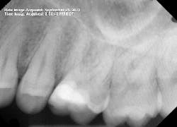
On a late Friday afternoon, one of our dental assistants told me she had been in severe pain for the past four days. No. 14 had a deep filling that was replaced last March, and now her tooth was causing constant pain. The tooth was extremely sensitive to touch, hot, and cold.
Diagnosis
Tooth No. 14 indeed turned out to be extremely sensitive to percussion. I recommended root canal treatment. We have a new associate in our practice, so I decided this would be a good procedure for her to observe. I ask all of the dentists in our office to review the article “Step-by-step procedure to simplified and efficient root canal techniques,”after which I like to demonstrate the techniques on a live patient.
Treatment
I started the root canal at 2:00 p.m. that Friday. The procedure was uneventful at first. I gained what I thought was great access, and then I started on instrumentation. Just as I began, my 0.10 TF file separated in the distobuccal (DB) canal. This was unusual since a file that large is relatively strong. It is mostly used to instrument the coronal third and rarely encounters any significant curvature.
I then successfully bypassed the file with a 15 c-file, but it separated as well. I decided there must be a hidden curve somewhere and would return to the DB canal after completing instrumentation on the remaining canals. Something interesting to note: The apex locator was not able to obtain a reading on the DB canal at any point.
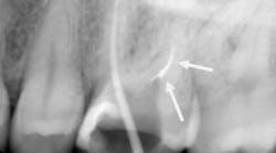
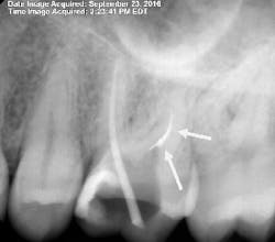
By 2:22 p.m., I was taking periapical x-rays with the gutta-percha to check my working lengths and evaluate the separated files. The x-ray showed that my access wasn’t as open as I originally thought it to be. The canal curved in two places, which caused the file separations. At this point I had two separated files—the 0.1 Twisted File (TF) file and the 15 mm hand file I had used to bypass the canal.
I proceeded to bypass the separated files with a 10 c-file. The apex locator still wasn’t giving me a reading. I was ready to obturate the canal, leaving all the extra metal as fill. Upon drying the canal with paper points, I noticed the paper point disappeared immediately toward the buccal. I took a step back to remove the buccal wall for better access. At this point, I was able to visualize the DB canal much better and was able to remove both of the separated instruments.
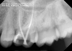
After obturation, it became apparent why the apex locator had given no reading for the DB canal. The foramen was located 3–4 mm away from the apex and on a curve that no file could reach. As a result of copious irrigation, the sealer was expressed from the apex.
Dentistry is a very humbling profession. But that’s what makes it exciting and interesting as well. After thousands of root canals, you still find one that surprises, teaches, and amazes.
ALSO BY DR. PETER MANN ...
A cost-effective method of creating a dental implant surgical guide for ridge augmentation
Interdisciplinary management of a complex maxillary implant restoration
Dental implants: The importance of patient compliance with immediate loading
Creating ideal cosmetic results for smile reconstruction: A tribute to Dr. Harvey F. Perlow
Narrow diameter dental implants in the esthetic zone


