When to do a functional analysis
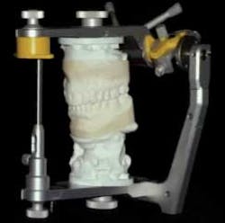
We have all had patients in our practice that present with very complicated cases including broken teeth, excessive wear, and gingival discrepancies, that are eager to learn how they can fix these issues. It isn’t uncommon for us to look at these patients and have no idea how we can predictably restore the esthetics while managing the occlusion.RELATED |Using the open tray technique when impressing implant sites Obviously we can’t tell our patients that we’re not sure how to proceed, so what do we typically do? We suggest that we need to take impressions and get some diagnostic casts that we will then study to learn how to best manage their case.RELATED | Close the access opening of implant abutments The problem is that if we just take the diagnostic impressions and use them as unmounted casts, we will actually learn very little about how to manage the occlusion and treatment. In order to really get as much out of the diagnostic models as possible, the models need to be mounted on an articulator using a facebow and centric relation bite. The diagnostic models can then be used for a functional analysis to correlate our clinical findings with the mechanics of occlusion, as well as determine the feasibility of the occlusal corrections. Anytime we take a set of models to the articulator to mount, they will always be mounted in CR. This allows us to evaluate the occlusal contacts in all of the mandibular positions. If the models were mounted in any other position, we would not be able to observe the occlusal contacts that are present when the condyles seat. Although it gives us the option, it doesn’t mean that we will be restoring the patient in CR, it just allows us to know all the occlusal possibilities and the contacts that arise when the condyles are seated. We don’t need to do mounted models on every patient and it’s helpful to know when it is necessary to do so:Exam findings reveal pathologic/failing occlusion. Key indicators in this particular situation could be a patient that presents with fractured/worn teeth or muscle problems. Anytime we see these findings, we need to do a functional analysis to be able to figure out if the occlusion can be corrected and if it’s a factor in the clinical findings.Significant amount of restorative dentistry is planned. When a patient presents with an ailing/failing occlusion, we typically don’t want to rebuild the same occlusal scheme that is there. Having the mounted models will allow us to determine what changes can/need to be made in order to improve the occlusal relationship; this can be done by possibly equilibrating or performing a diagnostic wax-up. From this point we can fabricate our provisionals and proceed to our final restorations.When we are unsure about the expected occlusal outcome. If we’re planning on equilibrating the occlusion and are unsure about where the occlusion will end up, we can mount our models and perform a trial equilibration on the diagnostic casts. The models can also help us determine if the best treatment option: Is orthodontic treatment preferable? Will the patient need restoration? Can it be done with composite or will indirect restorations be required? If you are uncertain about the outcome, you can’t go wrong with taking the time to mount the diagnostic models and perform a functional analysis.Reprinted with permission from Spear Education.
